


Osteochondrosis is a chronic degenerative-dystrophic disease that develops under the influence of various factors. Initially, the pathological changes occur in the nucleus pulposus (inner part of the disc), and then they extend to the annulus (outer sheath of the disc) and other elements of the spinal motion segment (SDS). ). This can be a result of the body's natural aging process, or it can be caused by trauma, increased load on the spine, and other causes. In all cases, osteonecrosis is only the first stage of disc destruction, and if left untreated, protrusions and hernias form, often requiring surgical removal.
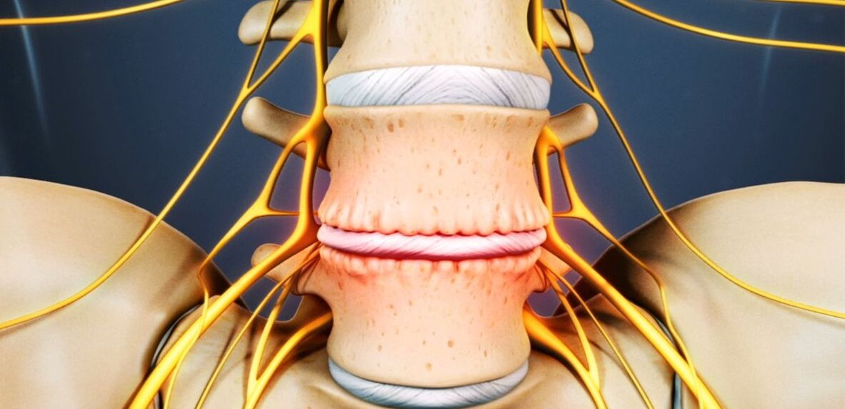
The disc is the cartilage formation that separates the vertebral bodies and acts as a shock absorber.
Osteochondrosis of the waist: what is it?
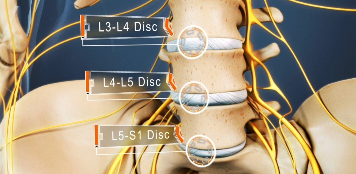
Between 48 and 52% of people have osteonecrosis. And osteonecrosis of the lumbar spine is the most common. This disease can affect any disc of the back spine, some or even all. Typically, disks L5-S1, L4-L5 suffer, less often than L3-L4. The upper lumbar discs (L3-L2 and L2-L1) are affected much less often.
The prevalence of lumbar osteonecrosis is due to the greatest load in performing any physical work, especially lifting and carrying weights, walking, running, sitting, and falls on the lower back. The lumbar spine consists of 5 vertebrae, they are much larger than the thoracic and cervical vertebrae. Accordingly, the discs separating them are larger in size. Normally, the lumbar region is slightly curved forward (physiological curvature). This is the last movable part of the spine and abuts the fixed sacrum, so most often they talk about osteochondrosis of the spine.
If previously osteonecrosis was considered an age-related disease, today its first manifestations can already be observed at the age of 15-19 years. Among the 30-year-olds, 1. 1% had severe symptoms of degenerative-dystrophic changes in the disc. And in representatives of the older age group (from 59 years old), clinical manifestations of the disease were already up to 82. 5%. At the same time, the incidence of the disease continues to increase steadily, which is mainly caused by the increase in the average age of the population of the country, but also by lifestyle changes that are not conducive to health.
Reason for development
Today, there is still no consensus on the etiology of degenerative spinal diseases. However, the main theory of their development is invisible. According to her, osteonecrosis is a consequence of previous damage to the discs and bony structures of the spine, as well as the appearance of inflammatory and other processes. The theory is that degenerative changes are genetically predetermined and, in fact, inevitable. And their clinical manifestations, especially in young and middle-aged people, are due to the influence of various endogenous and exogenous factors.
Thus, the development of osteonecrosis of the lumbar spine is facilitated by:
- hard manual labor, especially involving heavy lifting;
- sedentary lifestyle, sedentary;
- any back injury, including bruises;
- overweight;
- metabolic disorder;
- violation of posture, deformity of the back spine;
- Flat feet and other foot diseases;
- pregnancy, especially multiple pregnancies.

Pathogenesis
Regardless of the cause, disc degeneration occurs when the intensity of the catabolism (molecular cleavage and oxidation) processes of the matrix proteins begins to exceed the rate of their formation. One of the key points of this process is the undernutrition of the intervertebral discs.
Since they, like most adult cartilage, do not have a direct blood supply, since they have no blood vessels, the delivery of nutrients to them and the removal of metabolic products occurs bydiffuse with sequential compression and relaxation of the disc during movement. The main structure that powers the disc are the end plates located on its upper and lower surfaces.
In themselves, the endothelium is a double layer formed by cartilage and bone tissue cells. Accordingly, the cartilage abuts the disc, and the bone - with the vertebral body. They are distinguished by good enough permeability, which ensures intercellular metabolism, intervertebral disc matter, and passage of blood vessels in the vertebral bodies. Over the years, especially with the negative effects of external and internal factors, the structure of the intervertebral discs changes, the blood supply decreases, which leads to a decrease in the metabolic intensity in the discs. As a result, its ability to create new matrices is reduced, resulting in its density decreasing with age.
At the molecular level, this comes with:
- reduce the rate of diffusion of nutrients and metabolic products;
- reduced cell viability;
- accumulation of cell breakdown products and altered matrix molecules;
- decreased production of proteoglycans (the macromolecular compound responsible for the formation of new chondrocytes and the main source of chondroitin sulfate synthesis);
- collagen scaffold damage.
Possible consequences
As a result of these constant changes, the intervertebral disc becomes dehydrated and the nucleus pulposus loses its ability to adequately distribute the loads placed on it. As a result, the pressure inside the disc becomes uneven, and as a result the fibrous ring is overloaded and compressed in some places. Since this happens with every movement of a person, the circle is constantly subjected to mechanical stress. This leads to adverse changes in it.
In addition, often a decrease in disc height and elasticity leads to compensatory changes in adjacent vertebral bodies. Bone growths called osteoclasts form on their surface. They tend to increase in size over time and even merge together, ruling out the possibility of migration in the affected PDS.
Due to malnutrition causing damage to the collagen skeleton, under the influence of the pressure of the marrow nucleus at certain points, the normal structure of the fibers forming the annulus is broken. Without intervention, this will eventually lead to cracks and breaks in them. Gradually, more and more fibers of the annulus at the site of application of the pressure are torn, resulting in protrusion. This is especially facilitated when the load on the spine is increased. And since the lumbar region bears the primary load during movement and any physical activity, it suffers most often.
The protrusion of the intervertebral disc without the final rupture of the annulus and with its base size larger than the protrusion is called the condyle. When it ruptures completely in one place or another, a herniated disc is diagnosed.
With partial destruction of the annulus fibers, the pressure in the disc gradually decreases leading to a decrease in tension and the fibrous fibers themselves. This leads to a violation of its fixation and as a result pathological mobility of the affected spinal segment of motion.
The vertebral motor segment (SMS) is a structural and functional unit of the spine made up of intervertebral discs, adjacent vertebral bodies, and the joints, ligaments, and muscles attached to these bony structures.
Normal functioning of the spine is only possible with correct operation of the PDS.
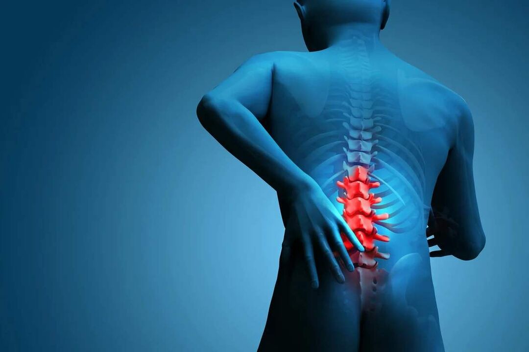
Symptoms of lumbar spine osteonecrosis
The disease may be asymptomatic for a long time, then begins to manifest as a slight discomfort in the lower back, which gradually worsens. But in some cases, osteonecrosis of the lumbar spine begins to be acute, immediately causing a strong pain syndrome. In most cases, signs of pathology appear for the first time after 35 years.
Back pain is the main symptom of the disease. It can vary in character, being both painful and dull, acute, continuous, or episodic. But basically for pathology, especially in the early stages of development, an alternation of periods of exacerbation and remission is characteristic, and both hypothermia or lifting a heavy object, orA sudden, unsuccessful movement can cause another deterioration in health.
Pain is often accompanied by numbness and tension in the back muscles. This condition is aggravated by exertion, sudden movements, heavy lifting, bending over, and even coughing and sneezing.
If, due to instability of the vertebral bodies, nerve roots extending from the spinal cord are clamped by one or another anatomical structure, this will lead to the development of appropriate neurological disorders. Their main manifestations are:
- shooting, severe pain radiating to the sacrum, buttocks, lower extremities, or perineum;
- sensitivity disorders of varying severity;
- limited mobility, limping;
- Weakness of the internal muscles due to pinched nerves.
In the lumbar spine, the spinal cord ends at the level of 1-2 vertebrae and enters the so-called cauda equina, which is formed by the accumulation of spinal roots. Moreover, each of them is responsible not only for the development of muscles, but also for specific organs of the small pelvis, so prolonged compression can cause disturbances in the work of therespective agency. This can lead to the development of impotence, infertility, gynecological diseases, hemorrhoids and other disorders.
The clinical picture of osteochondrosis of the lumbar spine, especially with a long course and the appearance of compression of the spinal roots, largely depends on the extent of the lesion, i. e. the specific discunderwent degenerative-dystrophic changes.
- L3-L4 disc failure - pain is given to the anterior-medial parts of the thigh, lower leg, and medial ankle. This is accompanied by decreased sensitivity of the anterior surface of the thigh, decreased severity or loss of knee jerking, as well as decreased quadriceps strength.
- L4-L5 intervertebral disc failure - pain is radiated from the upper part of the buttocks to the outer parts of the thighs and shins. Less commonly, this is accompanied by pain that radiates to the back of the foot, including 1-3 fingers. In these areas, there is decreased sensitivity and muscle weakness. Sometimes develop myasthenia gravis and incomplete extension of the big toe.
- L5-S1 intervertebral disc injury - pain that begins in the midsection of the buttocks and descends to the heel along the posterior surface or back of the thigh and lower leg and may capture the outer edge of the foot, like 4-5 toeshand. In these areas of the lower extremities, there is a decrease in sensitivity, and the stomach and gluteal regions often decrease in size, accompanied by their weakness. If spinal roots traverse at this level of PDS are affected, a decrease or loss of the Achilles reflex and vegetative muscles can be observed.
L1-L2 and L2-L3 discs are rarely affected.
The pain associated with the disease limits a person and significantly reduces their quality of life. Because they exist for a long time and often recur, if they do not appear often, this cannot help but affect the psycho-emotional state. As a result, more than half of the patients showed signs of chronic emotional stress, depressive disorder, etc. v.
Diagnose
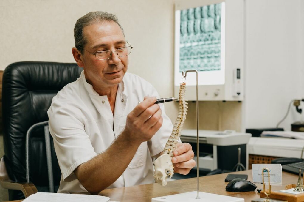
If there are signs of osteonecrosis of the lumbar spine, you should contact a neurologist or chiropractor. First of all, the doctor collects a history, which includes clarifying the nature of the complaints, the characteristics of the pain, the conditions of its occurrence and relief, the characteristics of a person's working life, etc. v.
The second stage of diagnosis, done as part of the initial consultation with the doctor, is the physical examination. During this procedure, the doctor will evaluate the condition of the skin, posture, depth of the physiological curves of the spine, the presence of its curvature, etc. v. The condition of the muscles around the spine, known as the paravertebral, must necessarily be evaluated, as they are often painful and overstressed, which is a response of the body to inflammation and the resulting pain.
On the basis of data obtained during examination and questioning of the patient, the neurologist may suspect the presence of osteonecrosis of the lumbar spine. But in order to rule out possible concomitant pathologies, as well as confirm the diagnosis and determine the exact extent of the lesion, the severity of degenerative-dystrophic changes in the intervertebral disc, and the associationof bone structure, laboratory and instrumental diagnostic methods are required.
Diagnostics in the laboratory
Different analyzes are not decisive in the diagnosis of osteonecrosis of the lumbar spine. They are aimed at assessing the extent of the inflammatory process and detecting concomitant disorders.
Therefore, they can be specified:
- UAC;
- HUG;
- blood test for sugar;
- blood chemistry.
Diagnostic tool
All patients with suspected lumbar osteonecrosis have:
- x-ray of the lumbar spine in two projections - allows you to determine the structure of the bone structure, detect abnormalities, bone formations, changes in aspects of the joints, etc. v. ;
- CT - allows you to detect changes in the bone structure at an earlier stage of development than x-rays, as well as identify indirect signs of osteonecrosis;
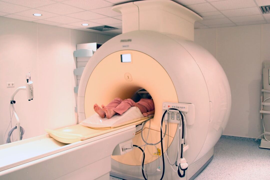
- MRI is the best method for diagnosing pathological changes in the formation of cartilage and other soft tissue structures, helping to detect the smallest changes in discs, ligaments, blood vessels, and spinal cord. accurately assess their severity and potential risk.
In addition, it may be recommended:
- densitometry - a method of determining bone density, helping to diagnose osteoporosis, which is especially popular in the elderly;
- myelography - allows you to assess the state of the cerebrospinal fluid passages of the spinal cord and the extent of damage to the protruding disc, which is especially important when there is a herniated disc that has formed in the lumbar spineback.
Treatment of lumbar osteonecrosis
When osteonecrosis is diagnosed, as a rule, all patients are initially treated conservatively, as long as there are no obvious and progressive neurological deficits. But her characters are chosen strictly individually.
Since the disease is chronic and the regenerative capacity of the discs is very limited, especially with pronounced degenerative-dystrophic changes, the main goals of therapy are to prevent their further progression and eliminate them. symptoms bother the patient. Therefore, the treatment is always complex and includes:
- drug treatment;
- manual therapy;
- physical therapy;
- exercise therapy.
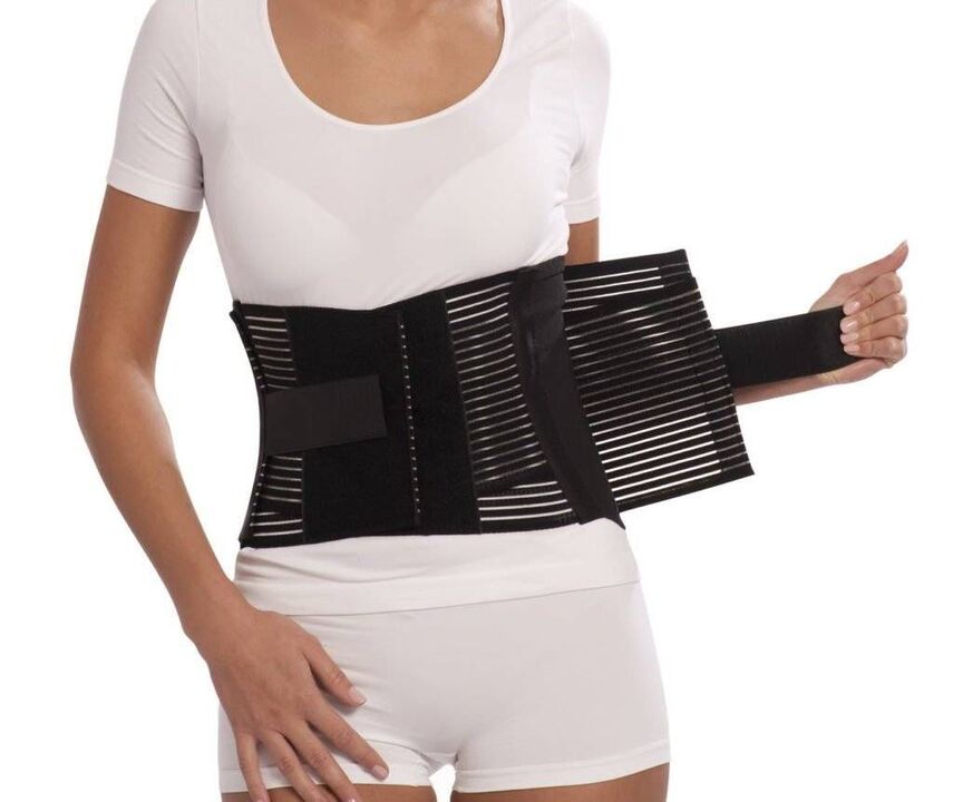
In the acute phase, the patient exhibits limited physical activity or even adherence to a bed rest regimen for 1-2 days. This will help relax the muscles and relieve pressure inside the disc. If you have to sit, walk, or do physical work for long periods of time, you should wear a corset to stabilize the lower back.
Conversely, after the end of the acute phase and during the remission phase of the disease, it is important to be as active as possible, but with caution and to rule out increased stress on the lower back. The patient will need to have the skills to sit properly, lift objects from the floor, carry heavy loads, as all these affect the course of the pathology. It is important to avoid sudden tilts and movements, lifting something from the floor or low surface, after bending the knees and not bending over. You should only sit upright on a chair that has a good back support effect. Also, during periods of sedentary work, it is important to take frequent breaks to exercise for short periods of time. It is important to avoid falls, jumping, running fast, and hypothermia.
With osteonecrosis, it is important to maintain body weight within optimal limits, and for obesity, a diet and physical exercise appropriate to the patient's condition are recommended. indicated, as excess weight puts an increased load on the lower back and causes a faster progression of pathological changes in the discs.
On average, conservative therapy is usually designed for 1-3 months, although it can last longer. But even after completing the main course prescribed by your doctor, it is still necessary to continue to take certain medications, exercise therapy, and follow lifestyle recommendations.
Medical therapy
The main components of drug therapy are drugs that are individually selected from the group of NSAIDs. When choosing them, the doctor takes into account not only the severity of the pain syndrome and the course of the inflammatory process, but also the nature of concomitant diseases, especially of the gastrointestinal tract, since NSAIDs if usedprolonged may adversely affect their condition. mucous membranes and cause exacerbations of various pathologies of the digestive system.
NSAIDs should be used for acute pain in the lower back and soon after it occurs. Preferably in 1-2 days. Depending on the severity of the patient's condition, they can be administered intramuscularly, as rectal suppositories, topical drugs, and in oral form. The admission period is no more than 2 weeks. In the future, a specially selected drug will be used as required, but try to avoid routine use.
Recently, more commonly preferred drugs, as an active ingredient, include selective cyclooxygenase-2 inhibitors.
In addition, patients are prescribed drugs of the following groups:
- muscle relaxants - help relax overstretched muscles and thus relieve back pain;
- chondroprotectors - improve metabolism in the intervertebral disc (especially effective when starting at the earliest stages of development of lumbar osteonecrosis);
- Vitamin B - contributes to the improvement of nerve conduction;
- antidepressants and anxiolytics - used for long-term osteonecrosis, which leads to depression, chronic fatigue and other psychological disorders.
With very severe pain, especially of neurological origin, blockade therapies are performed. They involve the introduction of anesthetics combined with corticosteroids into points near the pinched nerve, which helps to relieve pain quickly. But the procedure can only be performed in a medical facility by specially trained medical staff, because of the risk of complications.
Manual therapy
Manual therapy not only allows to improve the quality of blood circulation in the affected area, but also significantly reduces the severity and duration of pain of osteonecrosis. It effectively relieves muscle tension and allows you to remove functional blocks, which significantly increases mobility in the affected SMS.
In addition, through well-conducted manual therapy, it is not only possible to increase the distance between the vertebrae, returning them to their anatomical correct position, but also to release the compressed nerve roots. Thanks to this, the pain is quickly eliminated and the neurological disorders also disappear. It also reduces the likelihood of complications and disorders in the work of internal organs.
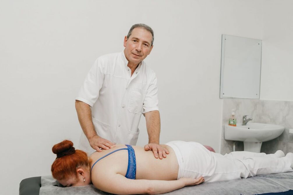
Additional positive properties of manual therapy are improved mood, enhanced immunity, activation of the body's natural recovery mechanisms and increased efficiency. Usually, after the first session, the state of health improves significantly, and in the future the effect will become more pronounced. As a rule, the course consists of 8-15 sessions, and it is important to complete it to the end, even if the state of health is completely normal.
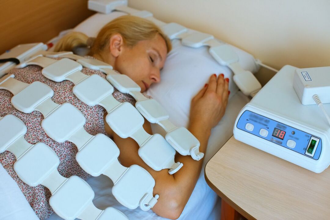
Physical therapy
After easing acute inflammation, physiotherapeutic courses are indicated, which not only relieve pain but also improve microcirculation, nutrition, and recovery in the area of degenerative-dystrophic changes. nursing. Usually, patients are prescribed:
- electrophoresis with the introduction of the drug;
- electrical nerve stimulation;
- therapeutic ultrasound;
- laser therapy;
- acupuncture therapy;
- UHF.
Which particular physiotherapy approach will work best, the frequency of administration, the duration of the course, and the likelihood of combinations with other types of exposure are determined individually for each patient.
Traction therapy gives very good results in the resorption of the lumbar spine. As a result, an increase in the distance between the vertebral bodies can be achieved, providing an immediate reduction in the load on the affected discs. After the session, to consolidate the results, the patient must wear an orthopedic bra.
exercise therapy
After eliminating acute pain, the treatment program is necessarily supplemented with exercise therapy. Its main job is to stretch the spine and relax the spasmodic muscles of the lower back. In addition, therapeutic exercises help strengthen the muscular corset, create reliable support for the spine and improve posture. During this process, blood circulation is definitely activated and metabolism improves, which has a beneficial effect on the nutrition of the intervertebral disc.
For each patient, a set of exercises was individually selected according to the degree of degenerative-dystrophic change, the patient's level of physical development, the nature of the co-morbidities, age andother factors. Initially, it is advisable to study under the guidance of an experienced therapist.
All patients with spondylolisthesis should go to the pool 2-3 times a week, because learning to swim minimizes the load on the spine, but allows you to effectively strengthen the back muscles.
Thus, lumbar spine tumor is one of the common diseases. At the same time, for a long time, it can deprive people of their ability to work, even leading to disability due to complications. Therefore, it is important not to ignore the first symptoms of the disease, when it is easiest to treat. With symptoms of pain, even numbness, limitation of movement, back pain, you need to contact a neurologist as soon as possible, make necessary examination and start treatment. In this case, it will be possible to end the pathological process and return to a full, normal life without significant pain and limitations.